Covering and lining cells
Begins in epithelial cells, which cover body surfaces and line many organs. Many common cancers are carcinomas.
A family of diseases
A cancer’s name usually starts with where it formed. The kind of cell involved, its microscopic appearance, and its molecular features add more detail.
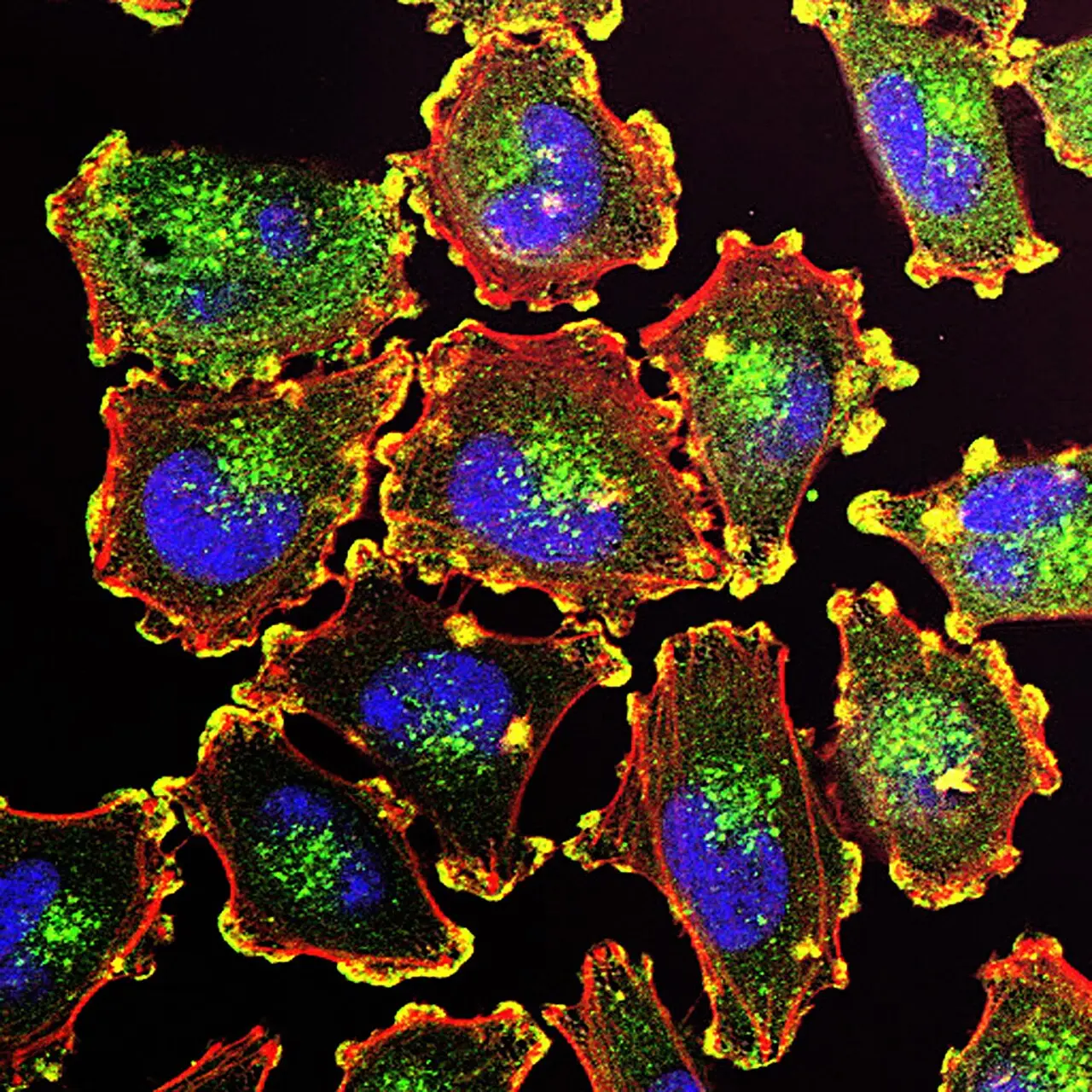
Melanoma begins in pigment-making melanocytes; fluorescent labels reveal selected structures.
NCI · Public domain ↗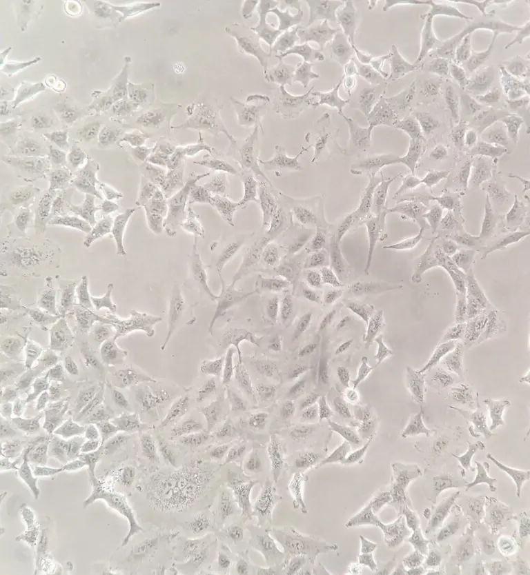
“Cancer” is an umbrella term. Classification narrows the story from a broad disease family to a particular origin, cell type, subtype, and set of features.
01 / THE STARTING POINT
Cancers are usually named for the organ or tissue where they first form. If cancer cells later spread elsewhere, the disease keeps the name of the original site. This is different from a new cancer that begins in the second location.
A cancer found in another part of the body may still be classified by where it began. That origin helps shape testing and treatment.
02 / CELL & TISSUE FAMILIES
Begins in epithelial cells, which cover body surfaces and line many organs. Many common cancers are carcinomas.
Begins in bone or soft tissues such as muscle, fat, blood vessels, or connective tissue.
Begins in blood-forming tissue such as bone marrow and generally does not form a solid tumor.
Begins in immune-system cells called lymphocytes. Hodgkin and non-Hodgkin lymphoma are broad groups with many subtypes.
Begins in plasma cells, immune cells that normally make antibodies.
Begins in pigment-making cells called melanocytes. Most melanomas form in skin, but they can begin in other pigmented tissues.
03 / CANCER MICROSCOPY GUIDES
Each visual guide connects licensed microscopy with plain-language explanations of cell origin, tissue pattern, pathology, and biomarkers. The images are educational examples, never tools for self-diagnosis.
Compare organized breast tissue with cancer histology, then learn how ER, PR, and HER2 add molecular context.
Open breast cancer guide →RESEARCH MODELExplore cultured A549 cells and learn why cell lines differ from diagnostic tissue.
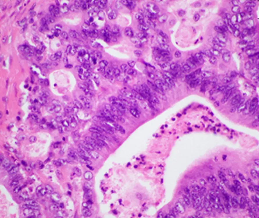
Open lung cancer guide →TISSUE ARCHITECTURECompare normal colorectal mucosa with adenocarcinoma and follow the route from screening to pathology.
Open colorectal cancer guide →TISSUE VS. CYTOLOGYDistinguish exocrine cancers, neuroendocrine tumors, and specimen methods.
Open pancreatic cancer guide →ELECTRON + FLUORESCENCESee how imaging technique changes what a melanoma cell reveals.
Open melanoma guide →03 / ORGAN & BODY SYSTEM
NCI organizes specific information by body location and system, including breast, lung, colorectal, pancreatic, prostate, ovarian, kidney, thyroid, brain and spinal cord, blood, bone, skin, and many others. Each site contains distinct types and subtypes; an organ name alone is not the complete diagnosis.
04 / WHY DETAILS MATTER
Microscopic pattern, grade, stage, and biomarkers can divide an organ-based cancer into more specific groups. Those differences can influence prognosis, which tests are useful, and which treatments are considered.
Pathology connects the organ of origin to a cell and tissue pattern.
Pathology & diagnosis →Biomarkers can reveal genes, proteins, and other features that are not visible by eye.
Genes & biomarkers →SOURCES / LEARN MORE
General education only. Classification and diagnosis must come from a qualified clinical team.