CELLWEAR Return home ↗
IMAGE RECORD / PUBLIC USEUPDATED SEPTEMBER 2, 2026

Trace every image

Rights should never be invisible.

Cellwear uses real scientific imagery only when its source and reuse status can be documented. The archive combines public-domain work with Creative Commons images, preserving the credit and license attached to every field.

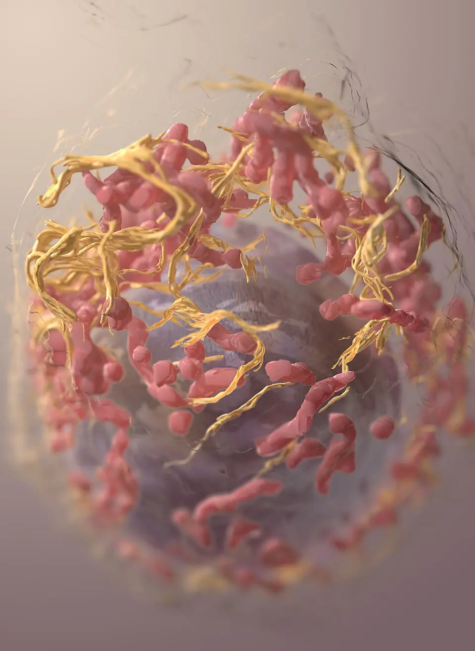
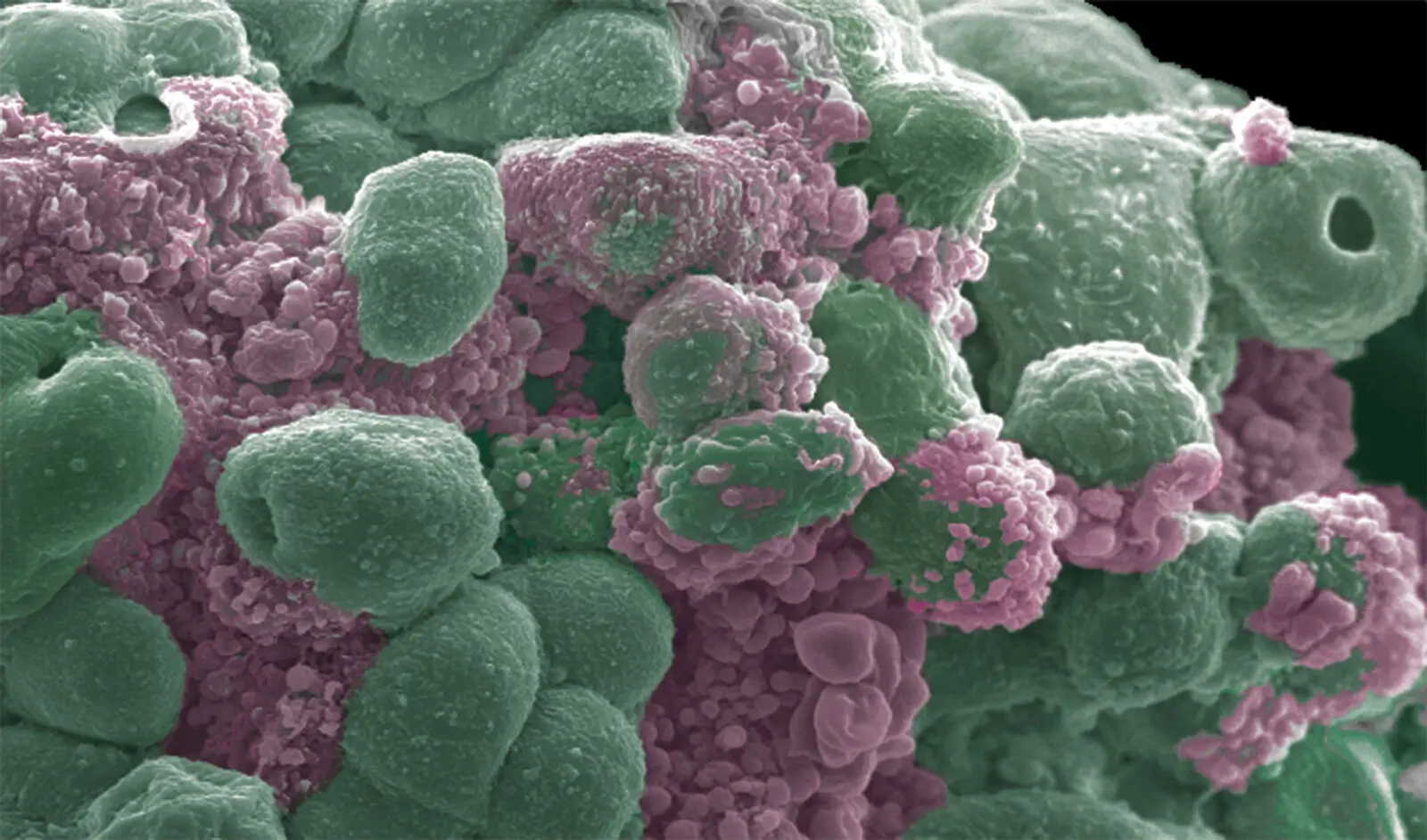
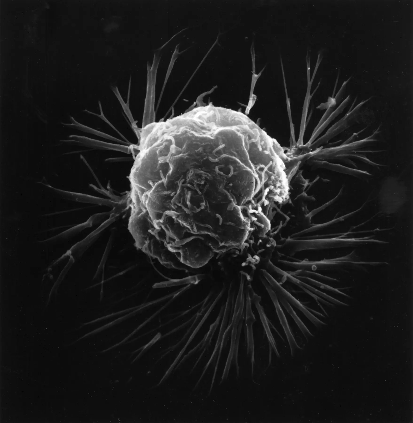
Scanning electron micrograph of a breast cancer cell
IMAGE 001 / PUBLIC DOMAIN

Breast cancer cell

A breast cancer cell photographed with a scanning electron microscope. The image shows the cell’s surface at high magnification; it should not be used by itself to identify or diagnose cancer.

Creator
Unknown photographer
Source
National Cancer Institute, 1980
Method
Scanning electron microscopy
Rights
Public domain; U.S. National Institutes of Health work
View the source and rights record
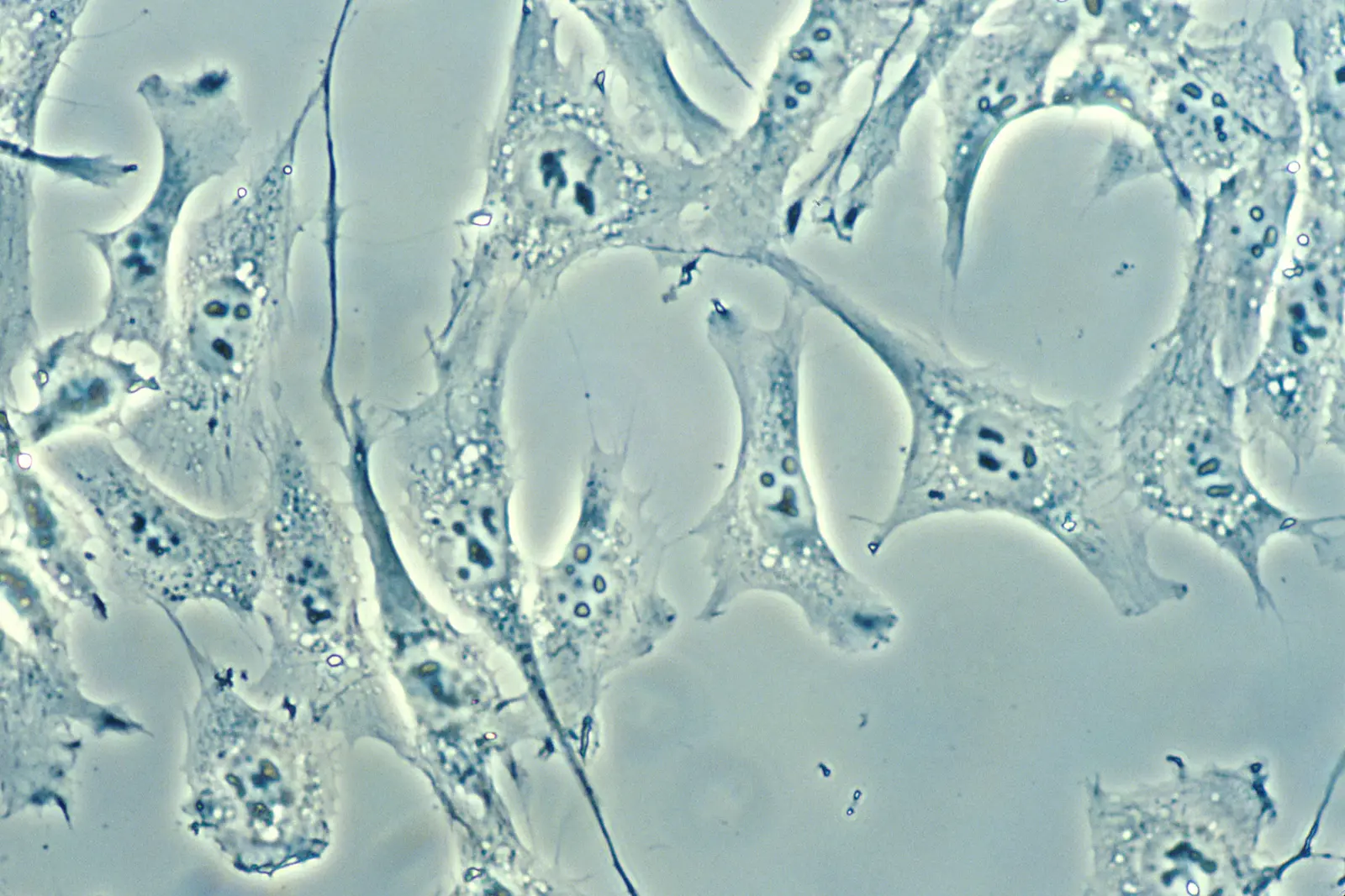
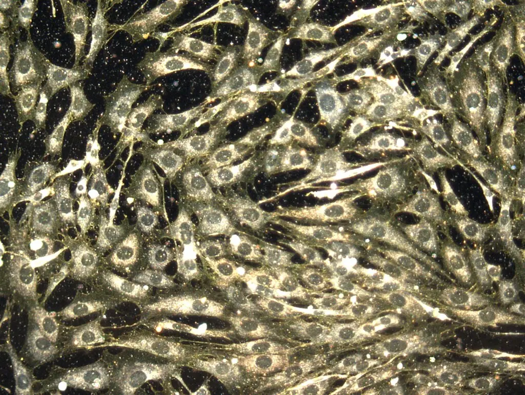
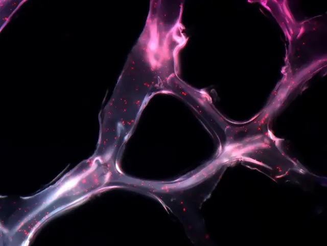
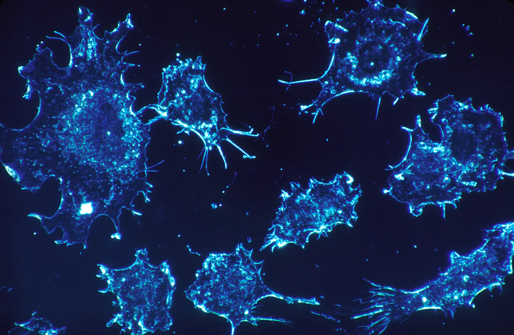
Cancer cells in culture illuminated using darkfield amplified contrast
IMAGE 002 / PUBLIC DOMAIN

Cancer cells in culture

Cancer cells cultured from human connective tissue, shown at 500× magnification using darkfield amplified contrast.

Creator
Dr. Cecil Fox
Source
National Cancer Institute, November 1987
Method
Darkfield amplified contrast
Rights
Public domain; dedicated by the photographer and identified as an NIH work
View the source and rights record
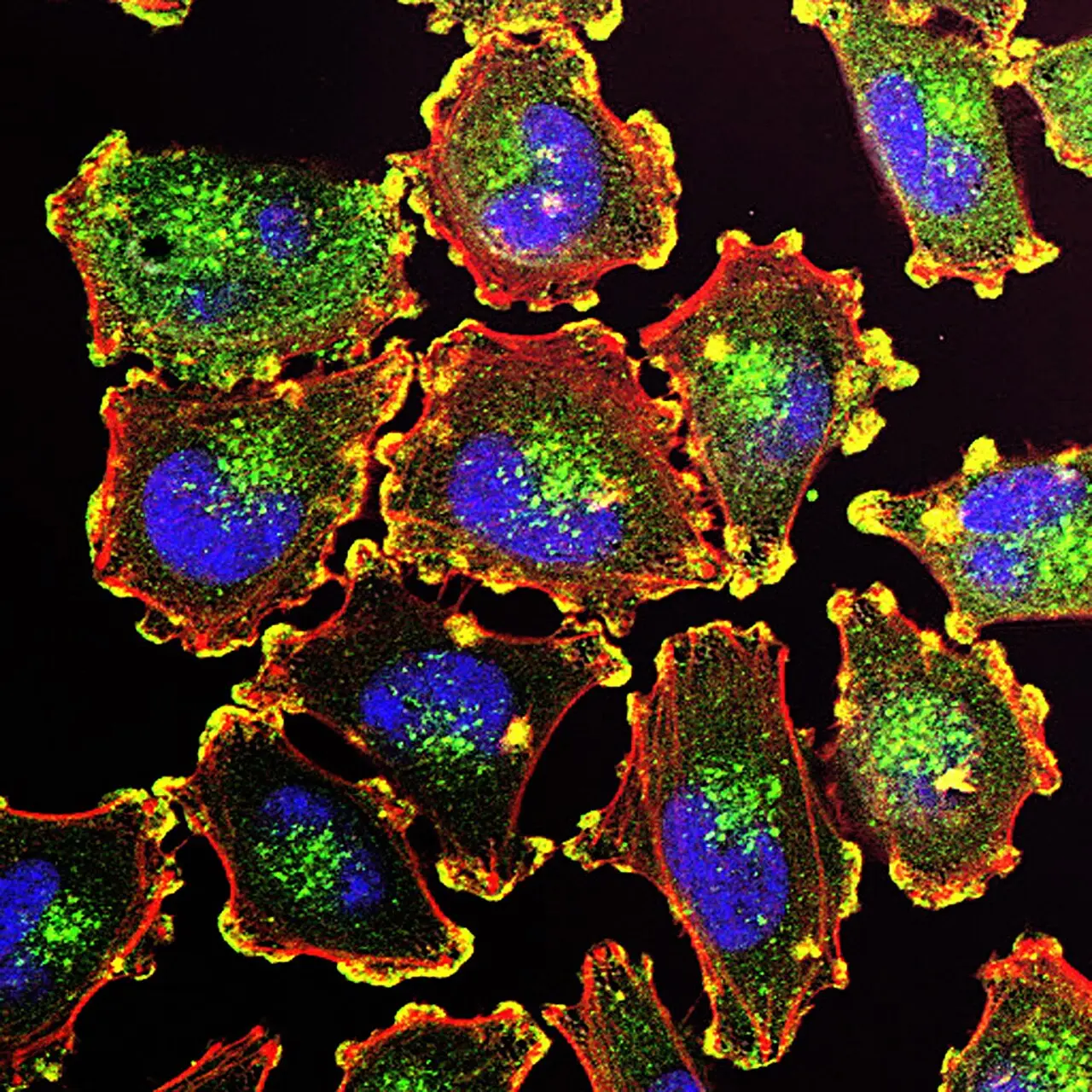
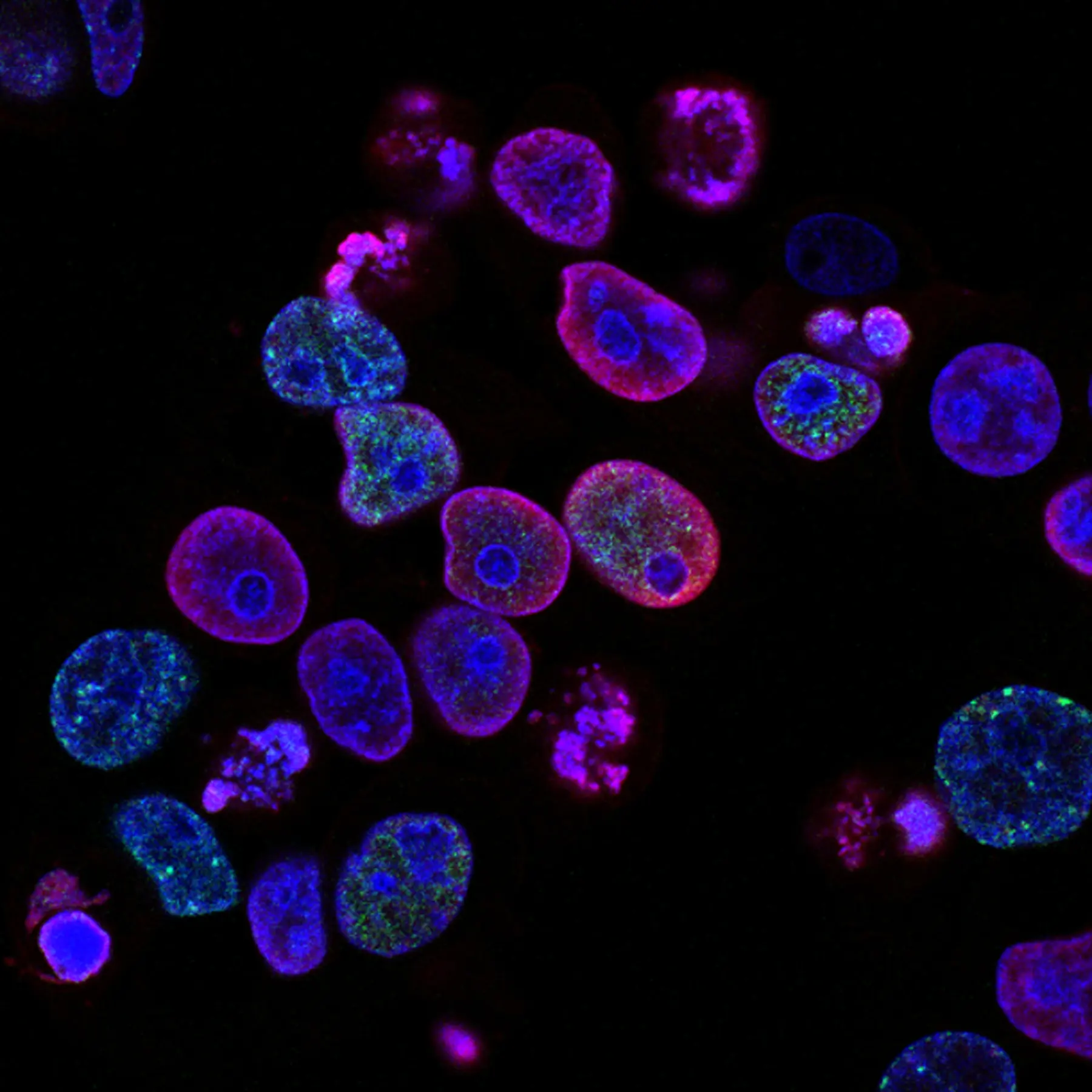
Fluorescent microscopy image of human colorectal cancer cells
IMAGE 003 / PUBLIC DOMAIN

Human colorectal cancer cells

Human colorectal cancer cells used in treatment research. Blue marks nuclei, while red and green markers highlight different cellular activity.

Creators
Yves Pommier and Rozenn Josse
Source
National Cancer Institute
Method
Fluorescence microscopy
Rights
Public domain; U.S. National Institutes of Health work
View the source and rights record

Gallery rights ledger

Seventeen more fields. Every source visible.

These records cover the additional images in the microscopy gallery. Follow any source link to inspect the original file page, full-resolution asset, and rights statement.

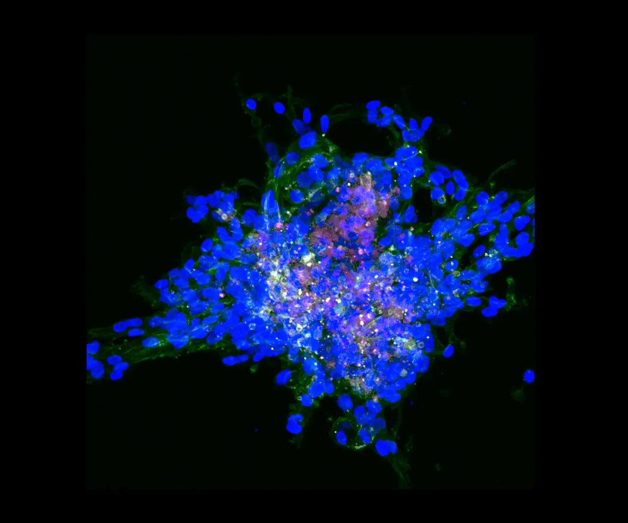
Three-dimensional fluorescence reconstruction of invasive breast cancer cells
IMAGE 008 / CC BY 4.0

Invasive breast cancer in 3D

Sshah74 · Wayne State University · 58 z-stacks, Zeiss LSM 780, 20×

Adapted by WebP conversion and resize.Source record ↗CC BY 4.0 ↗
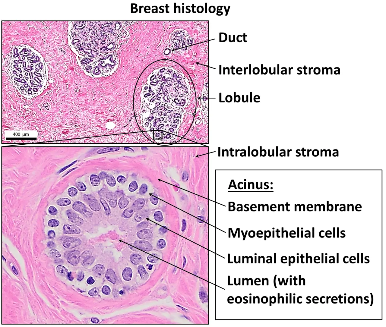
H and E stained normal breast tissue
IMAGE 009 / CC BY 4.0

Normal breast histology

Mikael Häggström, M.D.; source image contributors listed on the original record · H&E

Adapted by WebP conversion and resize.Source record ↗CC BY 4.0 ↗
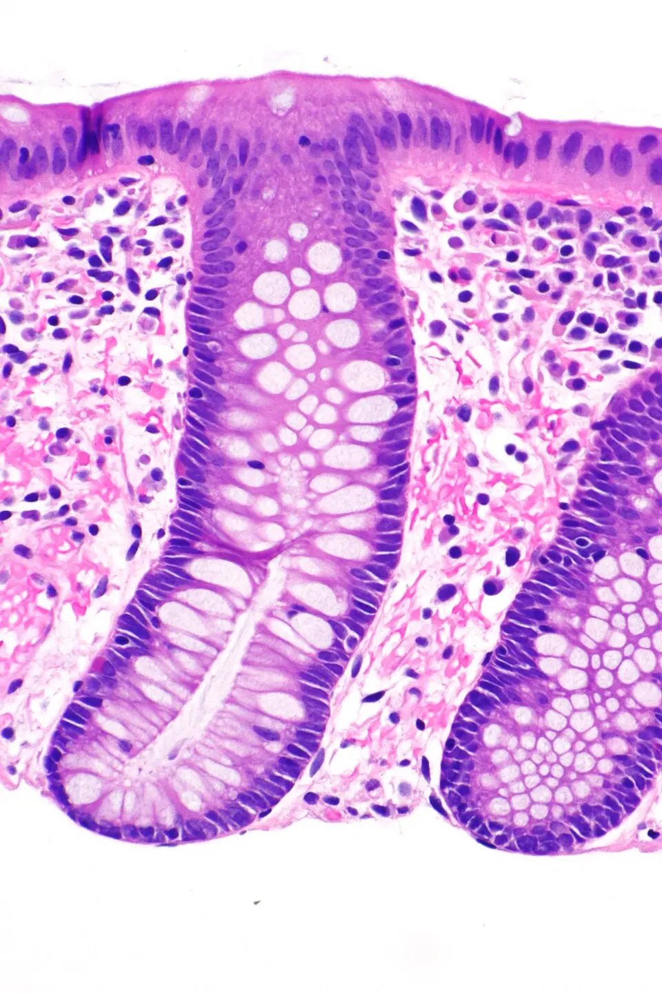
H and E stained normal colorectal mucosa
IMAGE 011 / CC BY-SA 3.0

Normal colorectal mucosa

Librepath · H&E biopsy

Adapted by WebP conversion and resize; this adapted file remains CC BY-SA 3.0.Source record ↗CC BY-SA 3.0 ↗
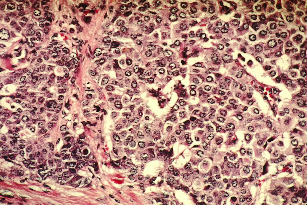
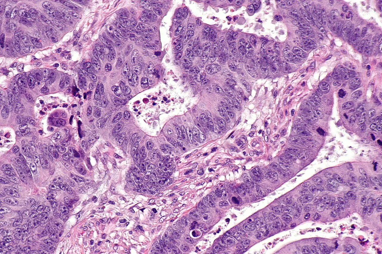
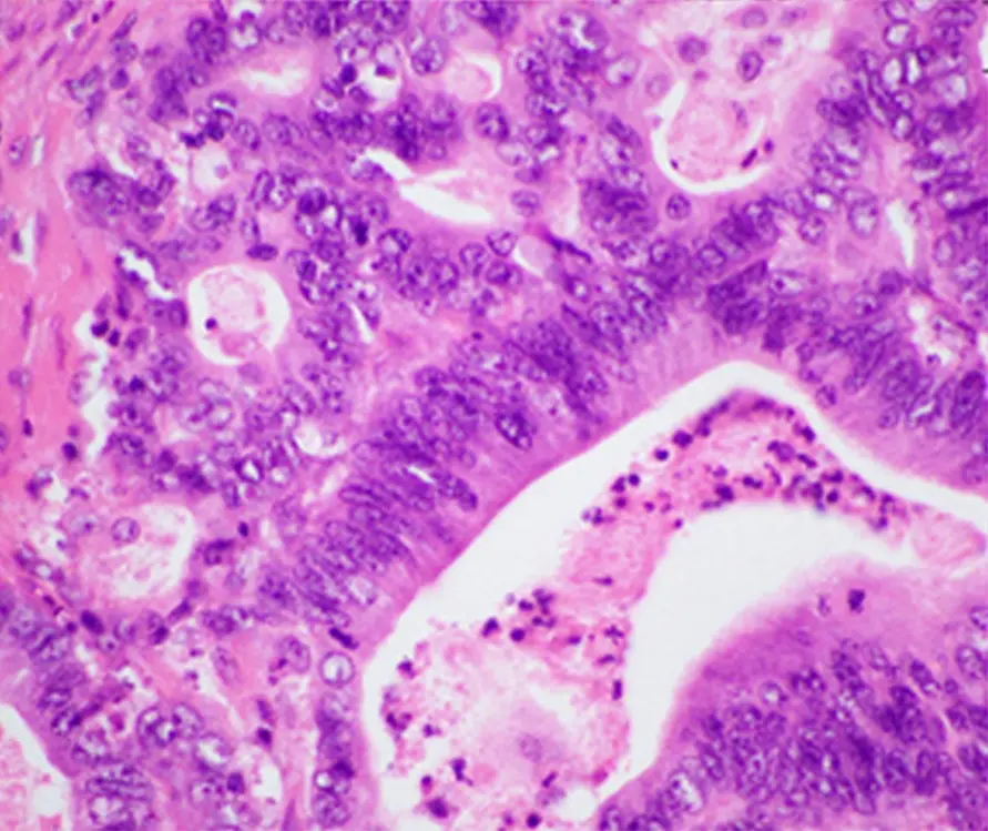
H and E stained colorectal adenocarcinoma
IMAGE 012 / CC BY-SA 3.0

Colorectal adenocarcinoma

Nephron · H&E histology

Adapted by WebP conversion and resize; this adapted file remains CC BY-SA 3.0.Source record ↗CC BY-SA 3.0 ↗
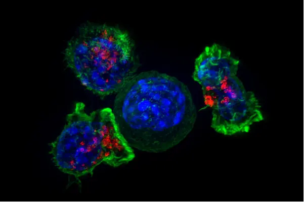
Fluorescence microscopy image of killer T cells surrounding a cancer cell
IMAGE 013 / PUBLIC DOMAIN

Killer T cells surround a cancer cell

Alex Ritter, Jennifer Lippincott-Schwartz and Gillian Griffiths · NIH · Fluorescence microscopy

WebP conversion and resize only.Source record ↗Public Domain Mark 1.0 ↗
Microscopy image showing gold nanoparticles within cultured cells
IMAGE 014 / PUBLIC DOMAIN

Gold nanoparticles in cell cytosol

Veronika Sapozhnikova, Konstantin Sokolov and Rebecca Richards-Kortum · NIH · Cancer nanotechnology research

WebP conversion and resize only.Source record ↗Public Domain Mark 1.0 ↗
Colorized electron microscopy image of prostate cancer cells exposed to nanosized drug carriers
IMAGE 015 / CC BY 4.0

Prostate cancer cells and nanocarriers

Khuloud T. Al-Jamal and Antonio Garcia · Wellcome Collection · Colorized scanning electron microscopy

Adapted by WebP conversion and resize.Source record ↗CC BY 4.0 ↗
Electron microscopy image of a branched artificial microRNA delivery scaffold
IMAGE 016 / CC BY 4.0

Artificial microRNA scaffold

João Condé, Nuria Oliva and Natalie Artzi · Wellcome Collection · Electron microscopy

Adapted by WebP conversion and resize.Source record ↗CC BY 4.0 ↗
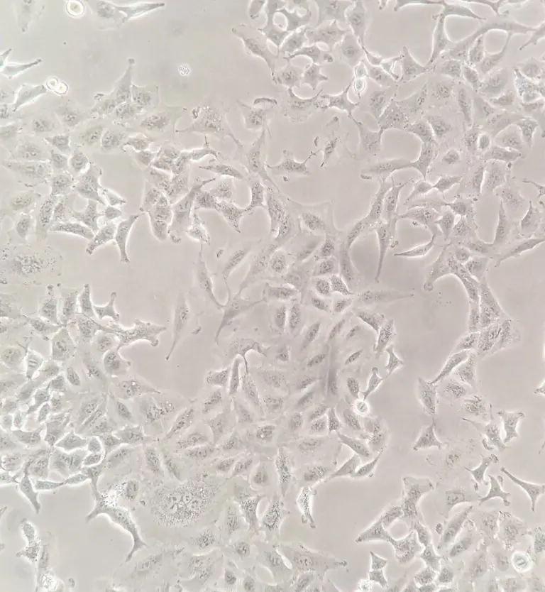
Brightfield microscopy image of cultured A549 human lung cancer cells
IMAGE 017 / CC BY 4.0

A549 lung cancer cells

Pinyapat Prisananuntakul · Brightfield microscopy · Cultured human lung cancer cell line

Adapted by WebP conversion and resize.Source record ↗CC BY 4.0 ↗
H and E stained histopathology image of pancreatic ductal adenocarcinoma
IMAGE 018 / CC BY 4.0

Pancreatic ductal adenocarcinoma

Pankaj K. Singh and coauthors · PLOS ONE · H&E histopathology

Adapted by WebP conversion and resize.Source record ↗CC BY 4.0 ↗
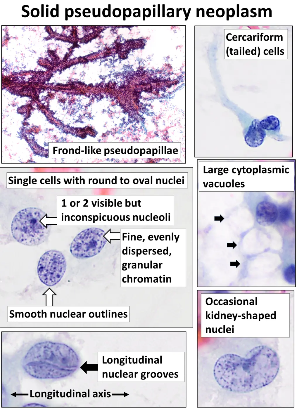
Labeled cytology panel showing a solid pseudopapillary neoplasm of the pancreas
IMAGE 019 / CC0

Solid pseudopapillary neoplasm cytology

Mikael Häggström, M.D. · Diff-Quik cytology · Labeled educational panel

WebP conversion and resize only.Source record ↗CC0 1.0 ↗
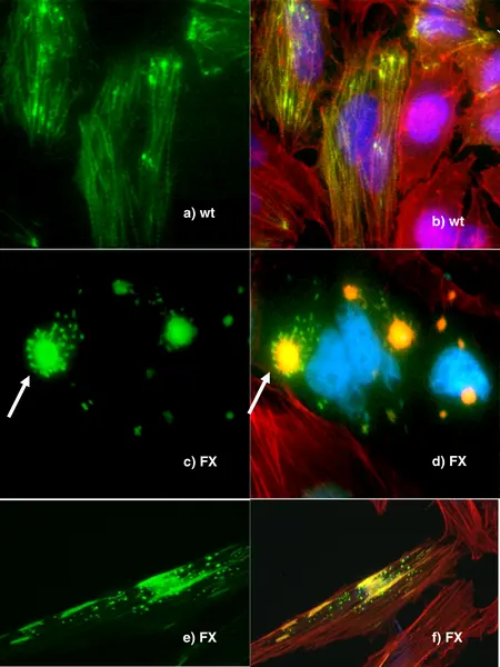
Fluorescence microscopy panels comparing palladin constructs in cultured HeLa cells
IMAGE 020 / CC BY 1.0

Palladin constructs in HeLa cells

Slater E, Amrillaeva V, Fendrich V, Bartsch D, Earl J, Vitone LJ, Neoptolemos JP and Greenhalf W · PLOS Medicine · Fluorescence microscopy

Adapted by WebP conversion and resize.Source record ↗CC BY 1.0 ↗

How the files were prepared

Optimized for the web. Not altered for meaning.

Cellwear converted source files to WebP, stripped metadata, and resized them for faster page loading. No labels, structures, or scientific interpretation were added. Creative Commons adaptations retain their original license, including ShareAlike where required. Product mockups are separate concept assets identified as microscopy-inspired art rather than medical imagery.