Paired field studies

Same tissue type. Different architecture.

These pairs place normal tissue beside cancer from the same general organ. They are separate specimens from different sources—not before-and-after images from one person—and their scale and preparation may differ.

PAIR 01 / CONNECTIVE TISSUE

The cleanest controlled comparison in the archive.

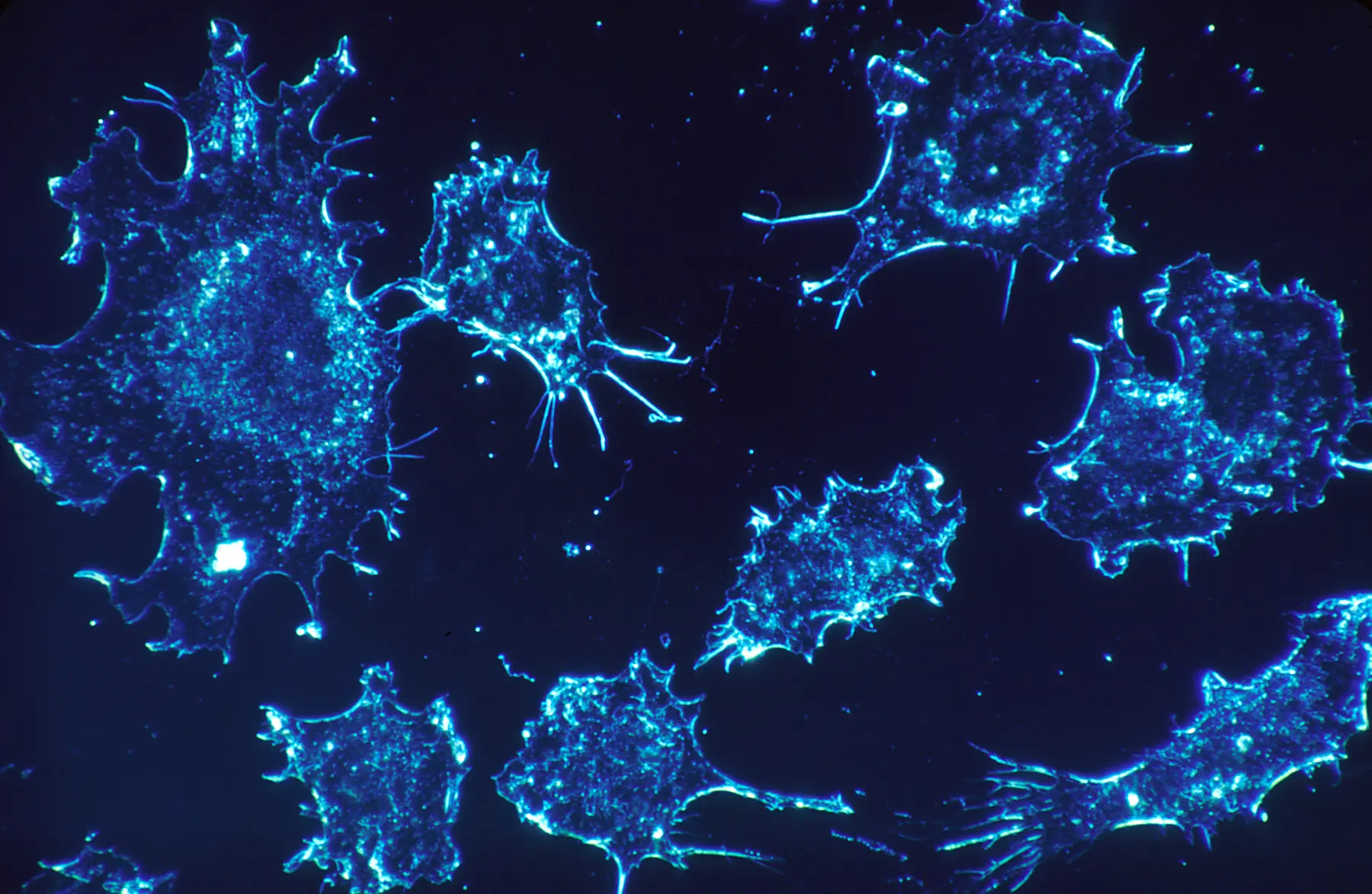
Both fields show cultured human connective-tissue cells photographed by Dr. Cecil Fox in November 1987 using darkfield amplified contrast at 500×. The normal cells and cancer cells are not documented as coming from the same donor or specimen.
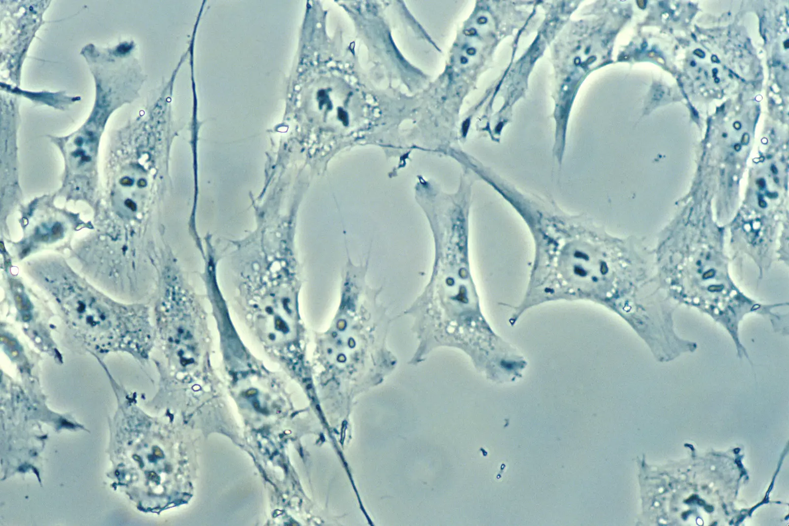
Normal human connective-tissue cells in culture using darkfield amplified contrastREFERENCE / NORMAL
Normal connective-tissue cellsDarkfield amplified contrast, 500× · Dr. Cecil Fox / NCIRights and context ↗
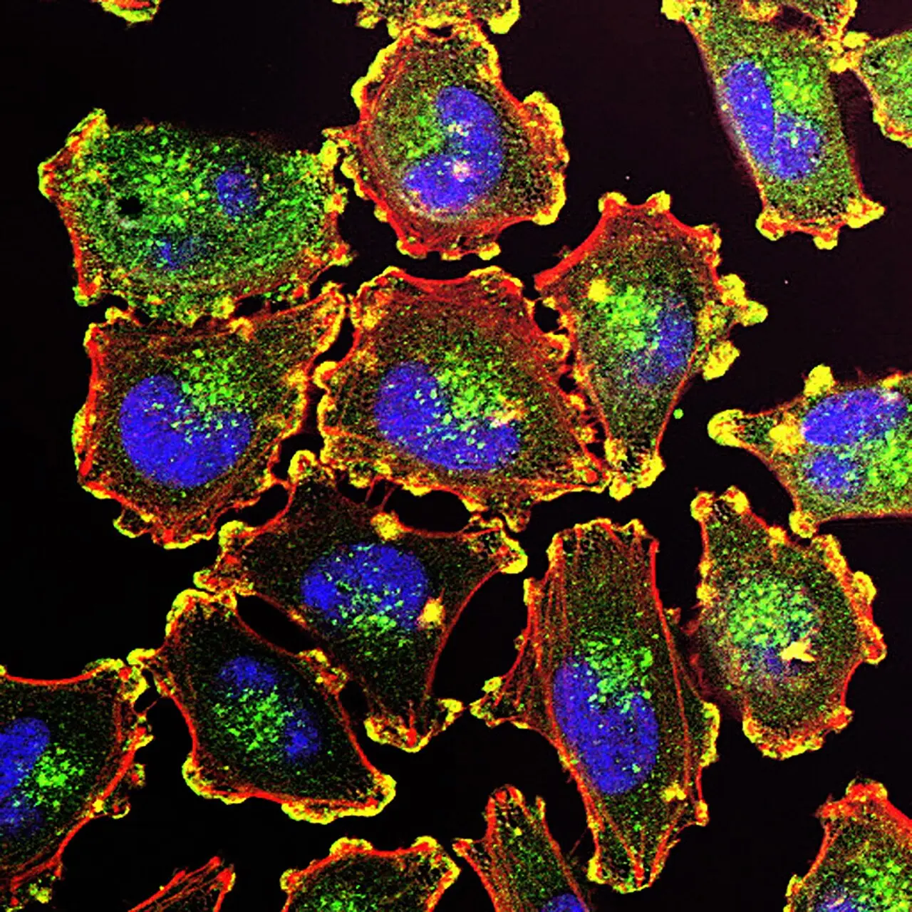
Cancer cells from human connective tissue in culture using darkfield amplified contrastFIELD / CANCER
Cancer cells from connective tissueDarkfield amplified contrast, 500× · Dr. Cecil Fox / NCIRights and context ↗
PAIR 02 / BREAST

Organized structures beside disrupted growth.

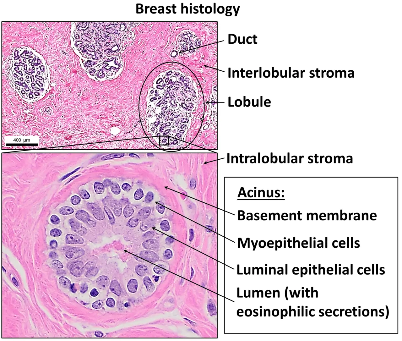
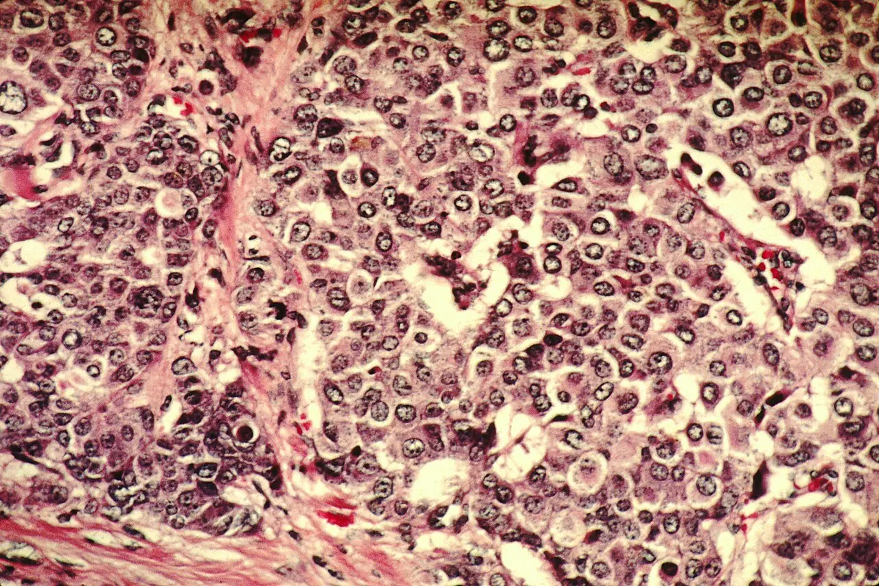
H&E staining commonly produces pink and purple tones. In normal breast tissue, cells form organized ducts and acini. The cancer image shows cells growing beyond their usual duct boundaries into surrounding tissue.

H and E stained normal breast tissue showing organized ducts, lobules, and an acinusREFERENCE / HEALTHY
Normal breast histologyH&E · Mikael Häggström, M.D. · CC BY 4.0Rights and context ↗
H and E stained breast cancer cells growing into surrounding tissueFIELD / CANCER
Breast cancer histologyH&E, 200× · Dr. Cecil Fox / NCI · Public domainRights and context ↗
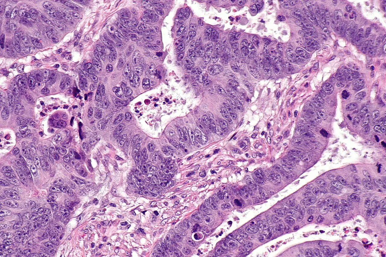
PAIR 03 / COLORECTAL

Orderly mucosa beside irregular glands.

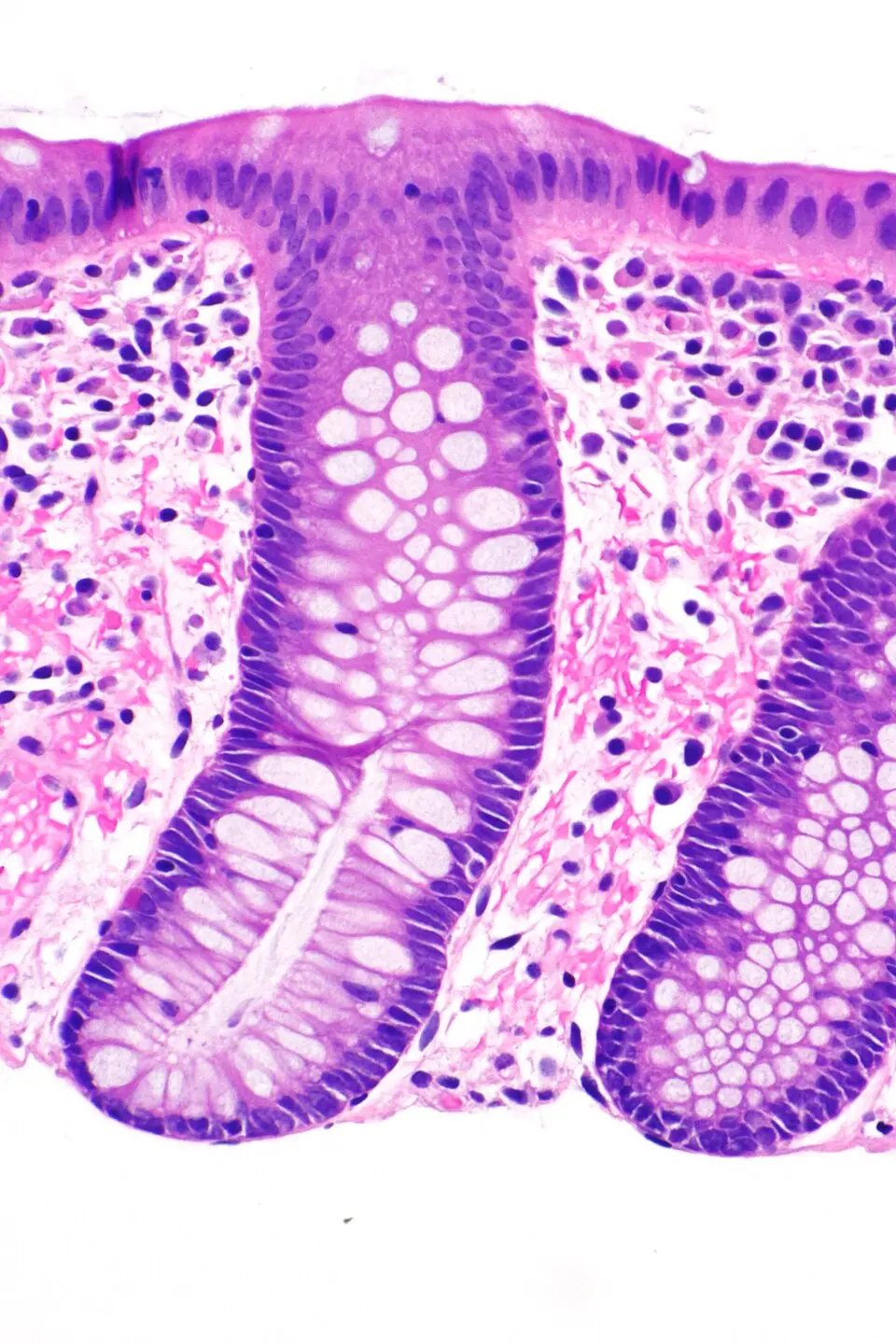
Normal colorectal mucosa forms repeated glandular structures with abundant mucus-producing goblet cells. In colorectal adenocarcinoma, gland shapes and the arrangement of cells can become irregular and crowded.

H and E stained normal colorectal mucosa at high magnificationREFERENCE / HEALTHY
Normal colorectal mucosaH&E biopsy · Librepath · CC BY-SA 3.0Rights and context ↗
H and E stained colorectal adenocarcinoma at high magnificationFIELD / CANCER
Colorectal adenocarcinomaH&E · Nephron · CC BY-SA 3.0Rights and context ↗

Look for patterns, not a self-diagnosis. Pathologists interpret complete specimens together with clinical and laboratory information. These comparisons are educational and cannot show whether any individual has cancer.

View the complete rights record