Biopsy or surgery
Cells or tissue are removed using a method suited to the location and clinical question.
Under the microscope
Cancer diagnosis can use physical exams, imaging, laboratory tests, and other procedures. In many cases, a biopsy and pathology report provide the evidence needed to confirm cancer and describe it.
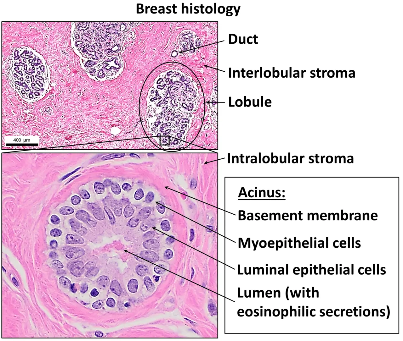
H&E staining helps make tissue architecture readable.
CC BY 4.0 · Rights ↗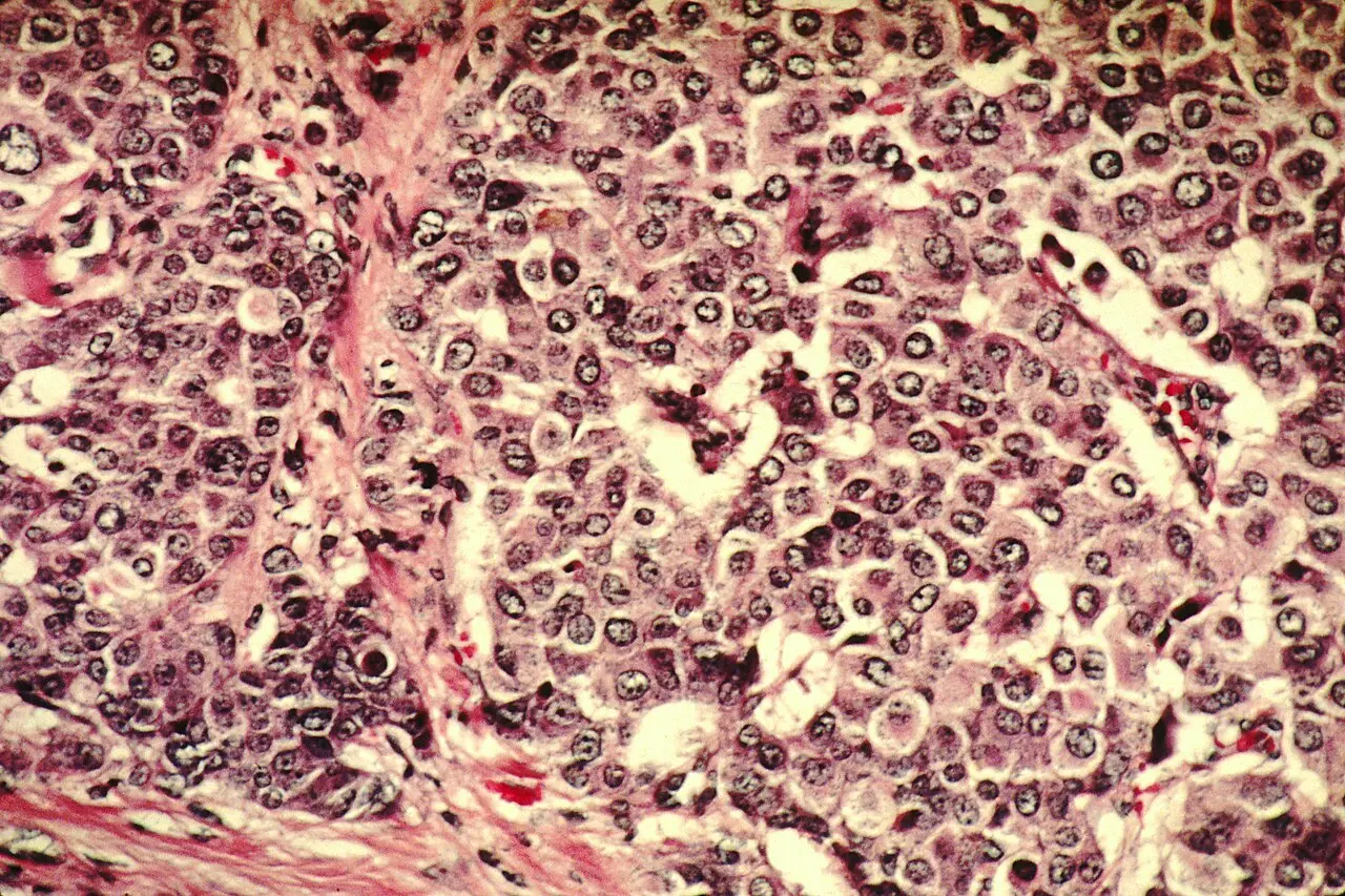
Pathologists assess patterns across a complete prepared specimen—not one isolated image.
NCI · Public domain ↗A microscope image is not a diagnosis by itself. Meaning comes from the sample, preparation method, expert interpretation, clinical context, and other tests.
01 / A PROCESS, NOT ONE TEST
A health professional may use a medical history, physical exam, laboratory tests, imaging, or procedures that collect cells or tissue. The exact route depends on what is being investigated and where it is located.
A biopsy is often needed to know for certain whether an area is cancer. The sample can also provide information about the type, grade, and biomarkers.
02 / FROM SAMPLE TO SLIDE
Cells or tissue are removed using a method suited to the location and clinical question.
The laboratory processes tissue into very thin sections. Stains create contrast between structures.
A pathologist evaluates cell shape, arrangement, and tissue architecture with the surrounding context.
The report communicates the diagnosis and other features to the clinical team.
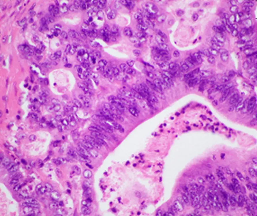
Histology preserves tissue relationships so architecture can be examined.
CC BY 4.0 · Rights ↗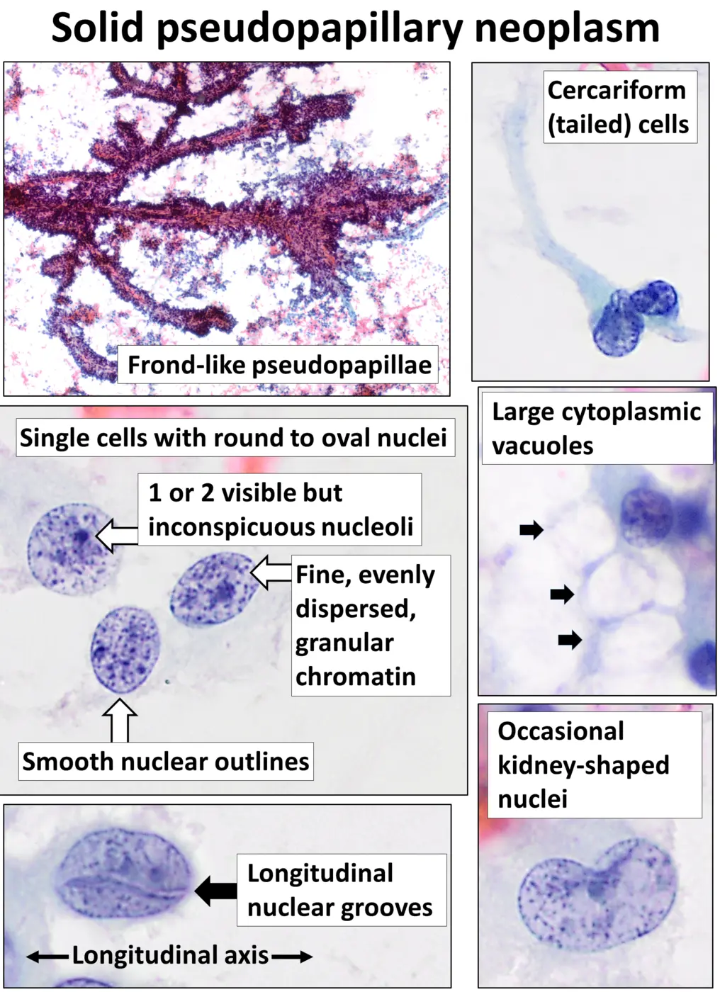
Cytology emphasizes individual cells. This is a different specimen and preparation—not a direct comparison with the histology image.
CC0 · Rights ↗03 / PATHOLOGY REPORT DECODER
Identifies the tissue, location, and often the procedure used to collect it.
Classifies the tumor based on the cells and tissue where it began.
Describes how much the tumor resembles or differs from normal tissue under the microscope.
For surgically removed tissue, records whether cancer cells are seen at a cut edge.
When nodes are sampled, the report records what was examined and what was observed.
Some reports include proteins, genes, or other features used in classification or care planning.
Not every report contains every section. Only the treating team can interpret a report in the context of an individual’s complete medical information.
04 / DO NOT MIX THESE UP
A pathologist determines tumor grade by examining how the cells and tissue compare with normal tissue. Stage describes how much cancer is present and whether it has reached nearby lymph nodes or other parts of the body. Systems differ by cancer type.
May help indicate how abnormal the tumor looks and how it could behave. The scale is cancer-specific.
Read the grade field note →Often considers the primary tumor, nearby lymph nodes, and whether cancer has spread farther.
Read the stage field note →SOURCES / LEARN MORE
General education only. This page cannot interpret a scan, biopsy, pathology report, or stage for an individual.