Non-small cell lung cancer
The more common group includes subtypes such as adenocarcinoma and squamous cell carcinoma.
Cancer cells under the microscope
The label “lung cancer” is a starting point. Microscopy, special stains, and molecular testing help identify the exact type and subtype.
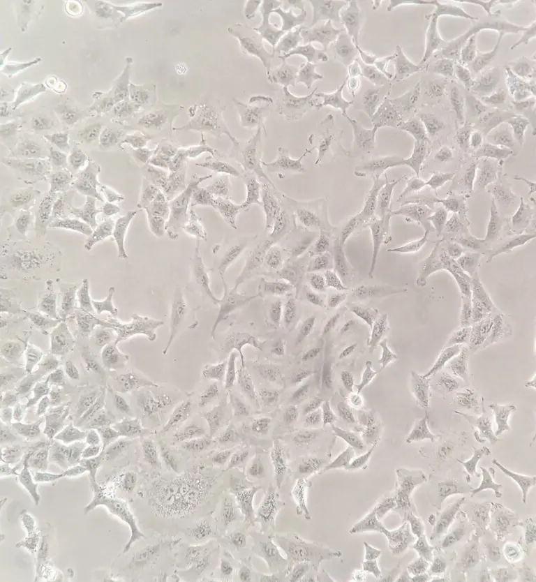
A549 cells are grown outside the body and used in laboratory research. They do not show the architecture of a patient’s lung tissue.
CC BY 4.0 · Image rights ↗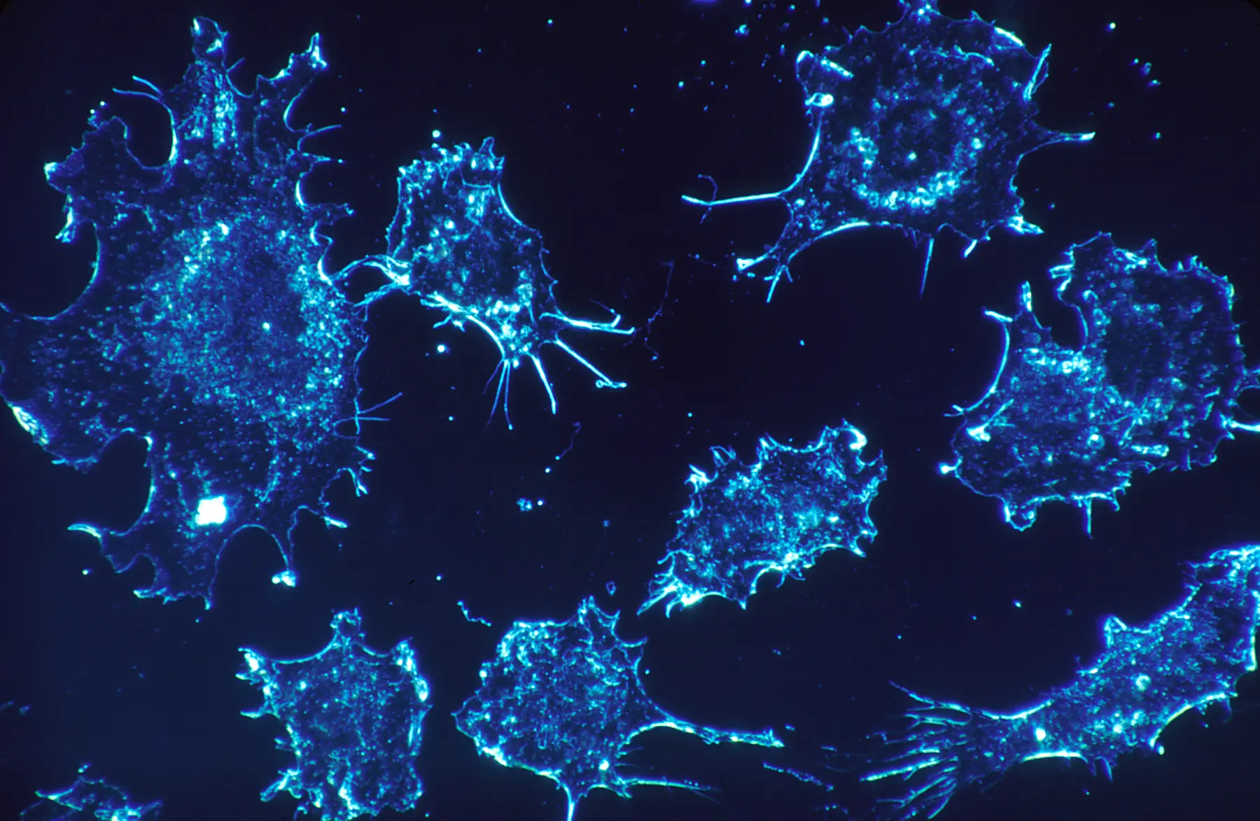
Color and contrast depend on the imaging method; they are not a cancer type by themselves.
NCI · Public domain ↗Lung cancer classification depends on the tumor’s microscopic and molecular identity. The result matters because different forms can behave differently and lead to different treatment options.
01 / RESEARCH VIEW
The A549 image above shows a human lung cancer cell line growing on a laboratory surface. Cell lines let researchers repeat experiments under controlled conditions, but they remove cells from the layered tissue, immune environment, blood vessels, and other context of a lung tumor.
A diagnostic pathology image would usually show cells within a patient tissue or cytology sample. This distinction is essential: a striking cellular image can represent research without being a view used to diagnose a particular person.
02 / CLASSIFICATION
The more common group includes subtypes such as adenocarcinoma and squamous cell carcinoma.
A distinct group named for its microscopic appearance and clinical behavior.
Those names are not interchangeable. A pathology report may include the specific subtype and level of certainty supported by the specimen. Not every lung cancer occurs in a person who smokes, and a single cause cannot be inferred from a microscopic image.
03 / ADDING EVIDENCE
A pathologist examines cell size, arrangement, nuclei, tissue pattern, and other features. Immunohistochemistry can use antibodies to detect selected proteins and help establish cell identity. For some non-small cell lung cancers, tumor biomarker testing can reveal molecular changes that may affect treatment planning.
Learn how staining and reports work in pathology and diagnosis, then explore what molecular tests can add in genes and biomarkers.
04 / LIMITS
Microscopy does not work as visual matching. Diagnosis requires the full specimen, laboratory preparation, clinical context, and often additional testing. Stage describes the extent of cancer in the body and cannot be determined from a close-up of cultured cells.
Ask what specimen, stain, scale, and method produced an image before deciding what it can teach.
SOURCES / REVIEWED 2026-09-02
General education only—not medical advice, diagnosis, or a substitute for professional care. Read our editorial standards.