Ductal and related cancers
Exocrine cancers are much more common. Pancreatic ductal adenocarcinoma is the best-known type.
Cancer under the microscope
The pancreas contains exocrine cells that support digestion and endocrine cells that produce hormones. Cancers from these systems are not the same disease.
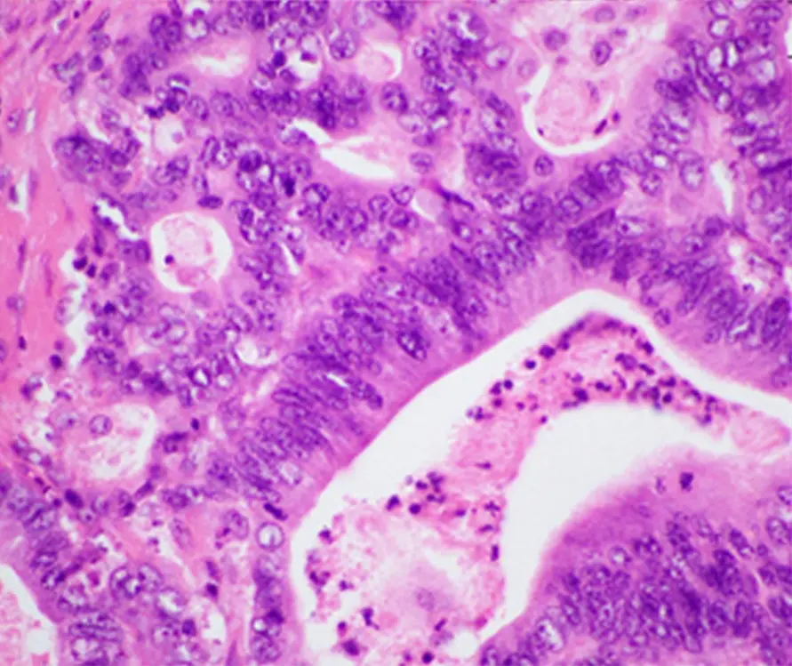
A tissue slice shows cells in relation to surrounding structures.
CC BY 4.0 · Image rights ↗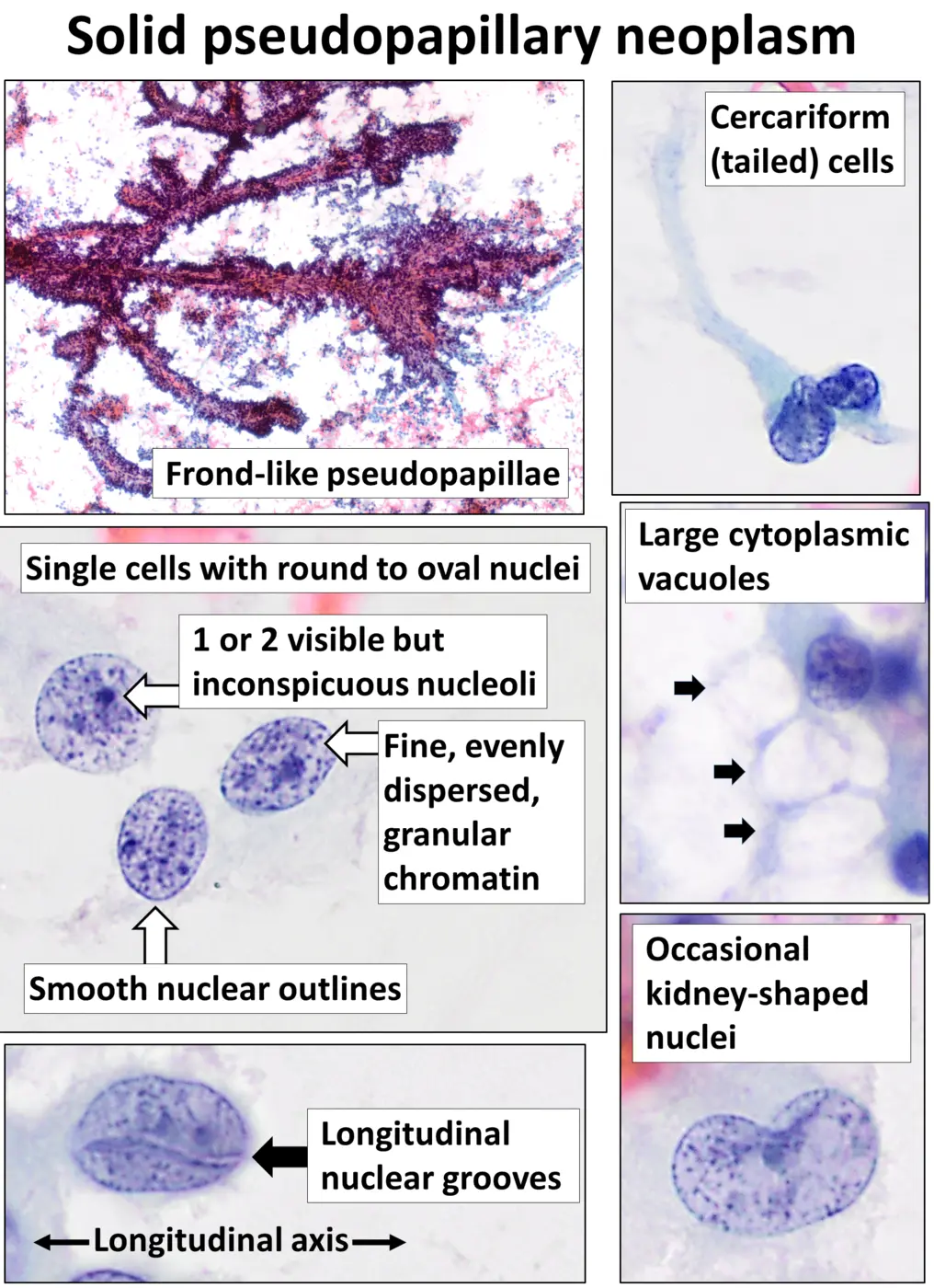
This is a different pancreatic tumor entity and a different specimen method.
CC0 · Image rights ↗“Pancreatic cancer” can describe tumors with different cells of origin and different behavior. The microscopic diagnosis establishes which disease is actually present.
01 / CELL OF ORIGIN
Most of the pancreas is made of exocrine tissue, which produces enzymes involved in digestion. Smaller groups of endocrine cells make hormones. Tumors arising from these systems are classified separately because their biology and treatment differ.
Exocrine cancers are much more common. Pancreatic ductal adenocarcinoma is the best-known type.
Pancreatic neuroendocrine tumors begin in hormone-producing cells and form a distinct group.
02 / DUCTAL ADENOCARCINOMA
Pancreatic ductal adenocarcinoma is an exocrine cancer. On a tissue section, pathologists assess gland formation, cell features, invasion, and the surrounding response. A small cropped image can introduce the pattern, but classification requires multiple fields and the full specimen.
03 / SPECIMEN CONTEXT
A tissue section preserves architecture: the relationship between tumor, ducts, supporting tissue, vessels, and other structures. Cytology emphasizes individual cells or small groups collected by a needle, brush, or fluid. Both can be useful, but they do not look identical and should never be treated as interchangeable images.
The image on the right is a solid pseudopapillary neoplasm—not pancreatic ductal adenocarcinoma. Similar location does not mean identical disease.
04 / BEYOND THE IMAGE
Microscopy identifies a tumor’s pattern and cell lineage. Grade describes differentiation and appearance; stage describes extent. Imaging, laboratory results, and sometimes molecular tests provide evidence that a microscope image alone cannot.
Continue with pathology and diagnosis or compare grade, stage, and biomarkers.
SOURCES / REVIEWED 2026-09-02
General education only—not medical advice, diagnosis, or a substitute for professional care. Read our editorial standards.