Cancer cells under the microscope
Breast cancer.
A tissue story with many subtypes.
Breast cancer is not one microscopic pattern or one biological disease. Tissue structure, cell features, and biomarkers work together to build a more precise diagnosis.
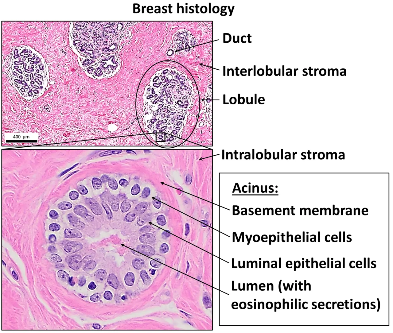
Normal histology provides a reference for the arrangement of ducts, lobules, and surrounding tissue.
CC BY 4.0 · Image rights ↗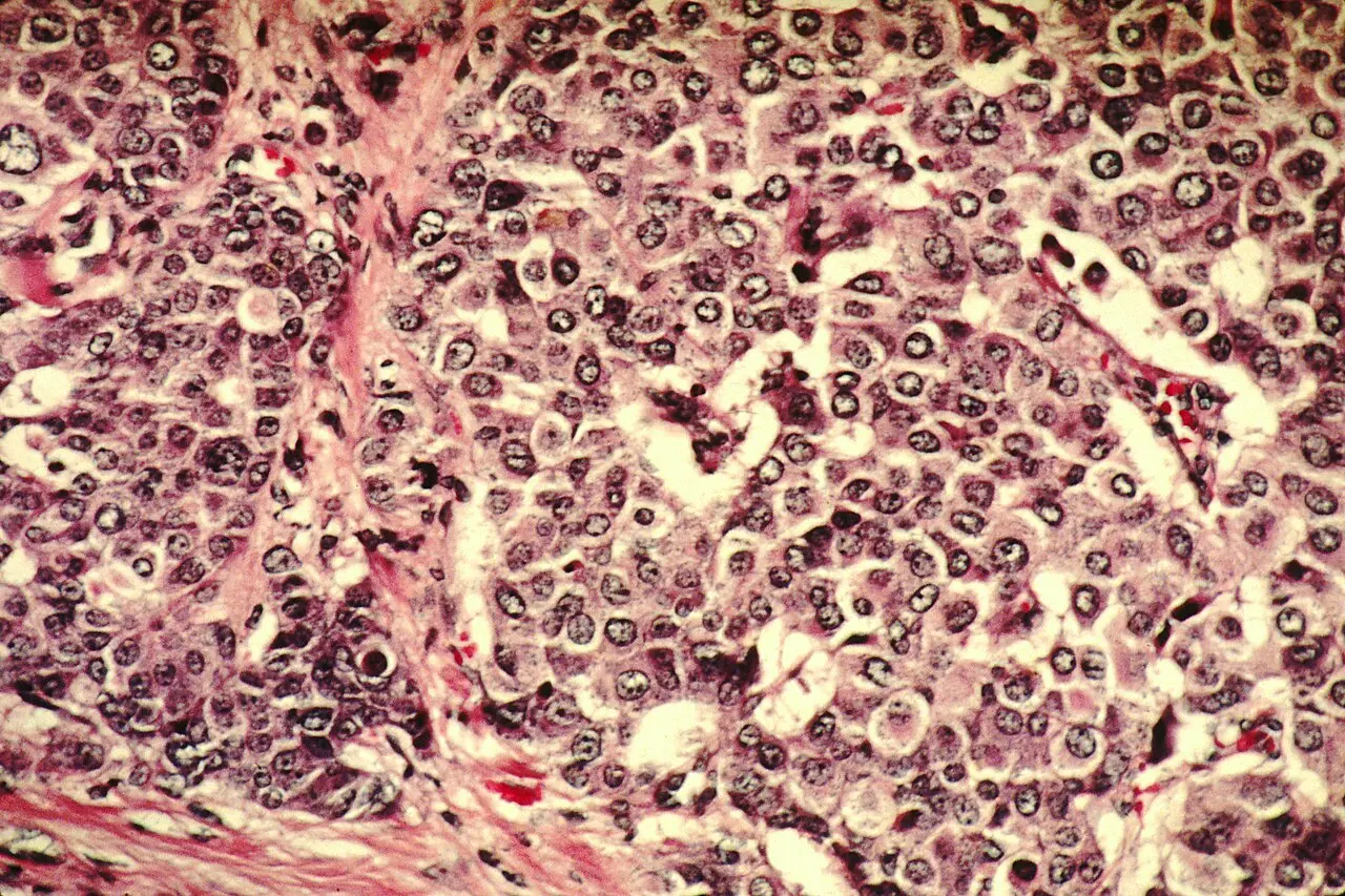
Pathologists examine patterns across the specimen, not one isolated cell or a web image.
NCI · Public domain ↗Under a microscope, breast tissue is read as an organized landscape. The key question is not simply “do these cells look unusual?” but how cells relate to ducts, lobules, boundaries, and one another.
01 / THE MICROSCOPIC VIEW
Pathologists read patterns across tissue.
A common stain called hematoxylin and eosin, or H&E, makes nuclei and surrounding tissue easier to distinguish. In healthy breast tissue, ducts and lobules generally retain an organized structure. In cancer, cells may become crowded, vary more in size and shape, and form patterns that disrupt or extend beyond expected boundaries.
Those visual clues help a pathologist identify whether a change is benign, in situ, or invasive and estimate grade. They are interpreted alongside the specimen’s location, additional stains, and clinical findings.
Microscopy examples teach visual concepts. Only a qualified pathology team examining a properly collected specimen can make a diagnosis.
02 / TYPE AND ORIGIN
“Breast cancer” names a family of diseases.
Many breast cancers begin in cells associated with ducts or lobules. A pathologist also determines whether abnormal cells remain within the structure where they began or have invaded nearby tissue. Ductal carcinoma in situ and invasive breast cancer therefore describe different findings, and invasive cancers include additional patterns.
Microscopic type matters, but it is only one layer. Tumor size, lymph-node findings, spread, grade, and molecular features add information used by the care team.
03 / MOLECULAR CLUES
ER, PR, and HER2 reveal biology the eye cannot.
Breast cancers are commonly tested for estrogen receptors (ER), progesterone receptors (PR), and the HER2 protein. These results help define groups such as hormone-receptor-positive, HER2-positive, and triple-negative breast cancer. The groups can behave differently and may respond to different treatments.
Biomarker results do not replace microscopy. They add another evidence layer to the pathology report. Learn how these tests fit into the larger picture in the cancer genetics and biomarkers guide.
04 / FROM SAMPLE TO REPORT
A biopsy turns a suspicious area into testable tissue.
Imaging can reveal an area that needs more investigation, but a biopsy provides cells or tissue for examination. A pathologist studies the sample, may order additional stains or biomarker tests, and reports the findings to the clinical team. Explore the complete route from specimen to report in pathology and cancer diagnosis.
SOURCES / REVIEWED 2026-09-02
Continue with authoritative information.
NCITypes of Breast Cancer↗NCIBreast Cancer Diagnosis↗NCIBreast Cancer Stages↗General education only—not medical advice, diagnosis, or a substitute for professional care. Read our editorial standards.