Scanning electron microscopy
Uses electrons to produce detailed surface information. The three-dimensional appearance is useful for research and visualization.
Cancer cells under the microscope
Melanoma begins in melanocytes, the cells that produce pigment. Electron and fluorescence microscopy can make their structure dramatic—but these research views answer different questions from routine pathology.
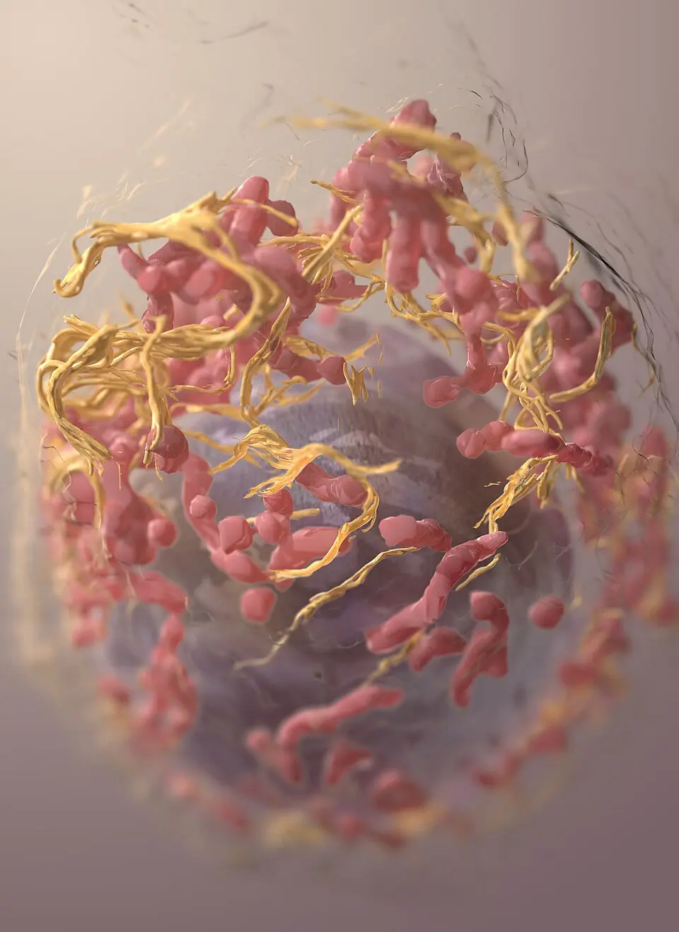
Scanning electron microscopy reveals detailed surface form in a research image.
NCI · Public domain ↗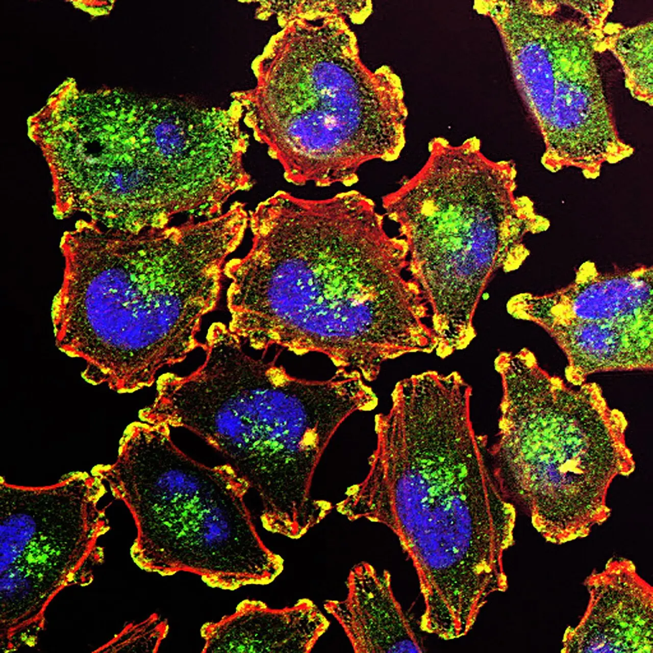
Fluorescent labels reveal specific molecules and structures—not natural cell color.
NCI · Public domain ↗Microscopy can reveal a melanoma cell’s surface or illuminate selected internal structures. Neither view alone establishes a diagnosis, stage, or treatment plan.
01 / CELL OF ORIGIN
Melanocytes make melanin and are best known for their role in skin color. Most melanomas arise in the skin, but melanocytes also exist in the eye and certain internal tissues, so melanoma can begin in other locations.
Melanoma is distinct from more common skin cancers that begin in other cell types. The name identifies its cellular origin, not simply the place where a spot appears.
02 / TWO LENSES
Uses electrons to produce detailed surface information. The three-dimensional appearance is useful for research and visualization.
Uses labels that emit light so researchers can track selected proteins, nuclei, or cell structures.
These colors and textures are produced by preparation and imaging choices. They should not be read as the natural appearance of a cell.
03 / ROUTINE DIAGNOSIS
For a suspicious skin lesion, a biopsy allows a pathologist to examine cells in their tissue context. The report may describe melanoma type, thickness, ulceration, mitotic activity, margins, and other findings depending on the specimen. Those details cannot be recovered from the research images above.
A close-up image online cannot confirm or rule out melanoma. A new or changing skin finding needs appropriate professional evaluation.
04 / MORE THAN APPEARANCE
Stage describes the extent of disease using clinical and pathology evidence. For some melanomas, biomarker testing can identify molecular changes that may affect treatment choices. These are different information layers—not competing answers.
Explore how they connect in grade, stage, and biomarkers and the cancer treatment atlas.
SOURCES / REVIEWED 2026-09-02
General education only—not medical advice, diagnosis, or a substitute for professional care. Read our editorial standards.