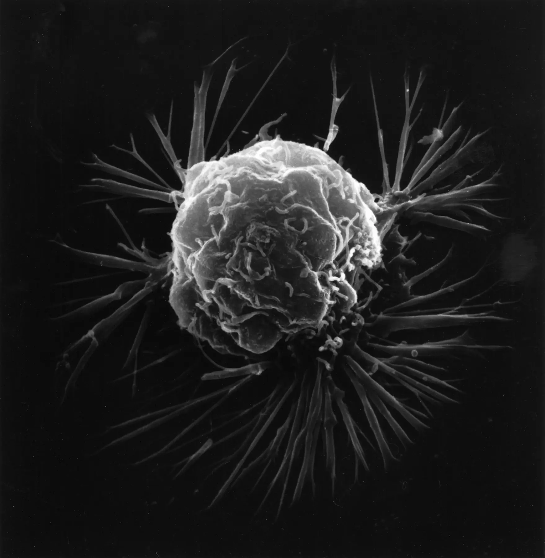
There is no single visual pattern called “cancer.” Cancer is a large family of diseases, and the appearance of a sample depends on where it came from, the kind of cells involved, how those cells are organized, and which tests were used to study them.
That complexity is exactly why microscopy matters. A pathologist—a doctor trained to identify disease by examining cells and tissues—combines visual findings with clinical information and, when needed, additional molecular tests. The result is a pathology report that helps establish a diagnosis and inform care.
01 / BEFORE THE MICROSCOPE
The image begins long before anyone looks through a lens.
A pathologist may examine tissue collected during a biopsy or surgery, or cells found in certain body fluids. For routine tissue examination, the sample is stabilized, processed, embedded in paraffin wax, and cut into extremely thin sections. Those sections are placed on glass slides.
The process matters because a microscope can only reveal useful detail when light can pass through a thin, well-prepared sample. Careful preparation also helps preserve the relationships between cells and the structures surrounding them.
A pathology image is not simply “a picture of cancer.” It is the visible result of sampling, preparation, staining, magnification, and expert interpretation.
02 / WHY COLOR APPEARS
Most tissue does not naturally arrive in violet and pink.
One of the most common staining methods is hematoxylin and eosin, usually shortened to H&E. According to the National Cancer Institute, hematoxylin makes genetic material and some structures inside the nucleus appear deep blue-purple. Eosin makes cytoplasm, collagen, connective tissue, and other supporting structures appear in shades of orange, pink, and red.
The stain creates contrast. It helps a pathologist distinguish nuclei from the material around them and see how cells relate to the architecture of the tissue. Other stains and tests can highlight different features, including specific proteins or molecular changes.
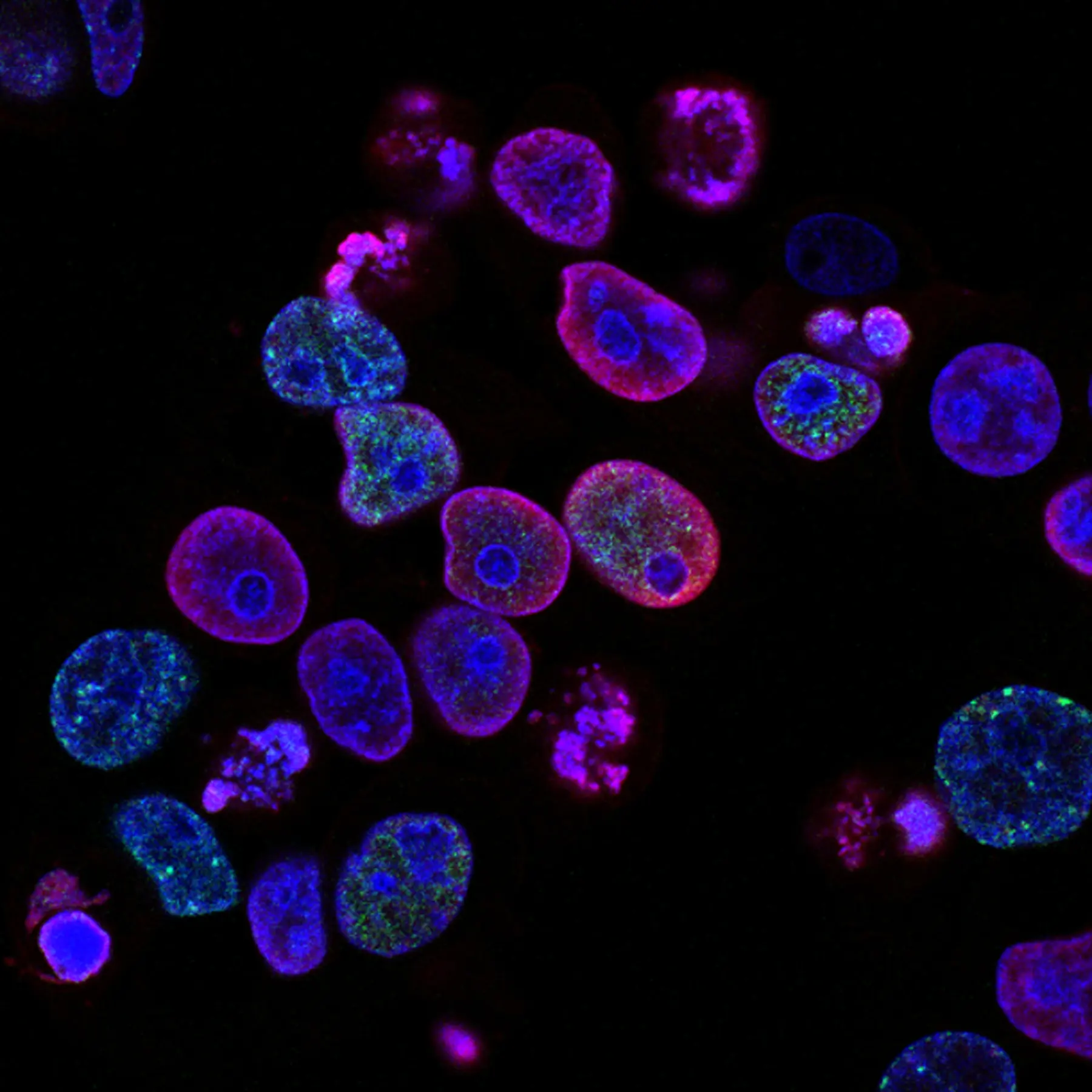
Human colorectal cancer cells with fluorescent markers. Yves Pommier and Rozenn Josse / National Cancer Institute; public domain. Rights and source →
03 / WHAT PATTERNS REVEAL
Pathologists read relationships, not isolated shapes.
A pathologist looks at far more than whether one cell appears unusual. The arrangement of cells, the structure of the tissue, the shapes and sizes of nuclei, and the boundaries between different areas can all contribute information.
The microscopic description in a pathology report may address the type of cells present, how abnormal they appear, how they are arranged, and whether certain features are seen in surgical margins or lymph nodes. Depending on the cancer, additional tests may look for proteins, chromosome changes, genes, or other biomarkers.
Cell and nucleus appearance
Size, shape, and visible organization can help distinguish patterns.
Tissue architecture
The way cells build glands, layers, clusters, or other structures can matter.
Margins and nearby tissue
Edges may help show whether abnormal cells reach a removed tissue margin.
Proteins and molecules
Additional testing can help refine a diagnosis or guide treatment decisions.
Grade is not stage.
Tumor grade describes how normal or abnormal cells look under a microscope. Stage describes the extent of a cancer, such as tumor size and whether it has spread. The systems used for both can vary by cancer type.
Read the NCI guide to tumor grade ↗04 / THE LIMITS OF AN IMAGE
One field of view never tells the whole story.
A single microscope image is a small view of a much larger specimen. It cannot by itself tell a person whether they have cancer, what treatment they need, or what their outcome will be.
Diagnosis may involve multiple areas of a sample, different magnifications, special stains, imaging, lab results, molecular tests, and clinical history. Even when a pathology report contains familiar words, their meaning depends on the exact cancer and the person’s complete medical context.
This is why Cellwear does not label artistic imagery as a diagnosis. When a design uses authentic scientific source material, it should identify the source, imaging or staining method when known, and educational context. When a design is interpretive, it should say so clearly.
05 / WHY WEAR THE IMAGE?
Awareness becomes stronger when curiosity has somewhere to go.
Traditional awareness symbols can show support, but they rarely explain the biology. Microscopic imagery creates a second layer: first a person notices the design, then they ask what it means.
That question is the opening. Clothing can connect a visual pattern to an article, a source, a glossary, or a story about a specific cancer type. The product becomes more than a symbol. It becomes an invitation to learn.
SOURCES / LEARN MORE
Continue with trusted cancer information.
NCIPathology Reports↗ NCITumor Grade↗ NCIHow Cancer Is Diagnosed↗This article provides general education and does not diagnose cancer, interpret individual pathology results, or replace guidance from a qualified health professional.